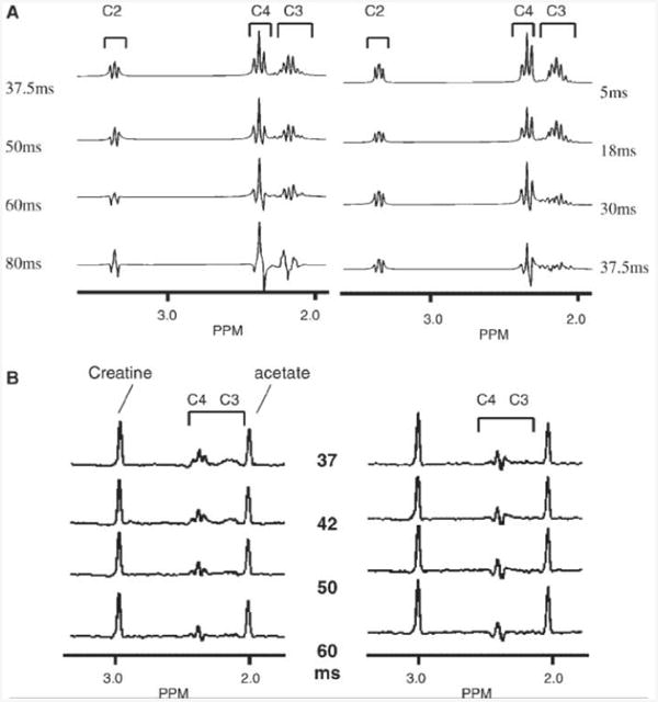
Figure 2.

(A) Simulation of two-dimensional LASER (left) and conventional double echo (right) sequences for glutamate, performed at 4T. (B) Phantom spectra (glutamate, creatine, acetate) showing retention of glutamate resonances, LASER (left), double spin echo (right). Timings are indicated, In both (A) and (B), the brackets indicate the individual glutamate resonances.
