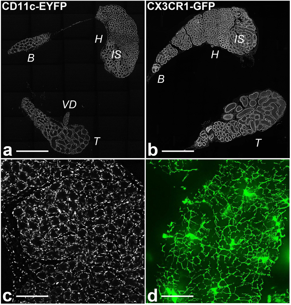
Figure 1.
The epididymis is densely populated by CD11c+ and CX3CR1+ cells. (a and b) “mosaic” pictures of whole CD11c-EYFP and CX3CR1-GFP mouse epididymis sections, respectively. IS: initial segments, H: head (caput), B: body (corpus), T: tail (cauda), VD: vas deferens. (c and d) higher magnification pictures of the initial segments, showing numerous CD11c-EYFP+ cells located at the periphery of the epididymal tubule. Bars = 2 mm (a and b), 250 µm (c) and 50 µm (d). High-resolution pictures for panels a and b are available online (online material S1 and S2).

