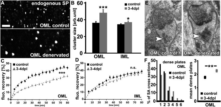
Fig. 2.
Denervation-induced homeostatic synaptic scaling is accompanied by structural changes of SP clusters and SA organelles. (A) Control and denervated wild-type cultures were stained for endogenous SP and mean cluster sizes were determined in the OML and IML. (Scale bar: 2 µm.) (B) A considerable increase in mean SP cluster sizes was detected in the OML following denervation. In the IML the effect was less pronounced (n = 6–10 cultures per group, three visual fields per culture). (C) Quantitative evaluation of GFP/Sp cluster fluorescent recovery after photobleaching (FRAP). Statistical comparisons performed with averaged values between 65–80 min per cluster (details given in SI Materials and Methods; n = 86 clusters from 12 control cultures; n = 70 clusters from 9 denervated cultures; six to nine clusters bleached per culture). (D) No significant difference in FRAP of GFP/SP clusters was observed in the IML of control and denervated cultures. (n = 54 clusters from eight control cultures; n = 52 clusters from seven denervated cultures; six to nine clusters bleached per culture). (E and F) The mean stack number of SAs was increased in the OML after denervation (n = 120–137 SAs in six cultures each; 20–25 SAs per culture). **P < 0.01; ***P < 0.001; NS, not significant.

