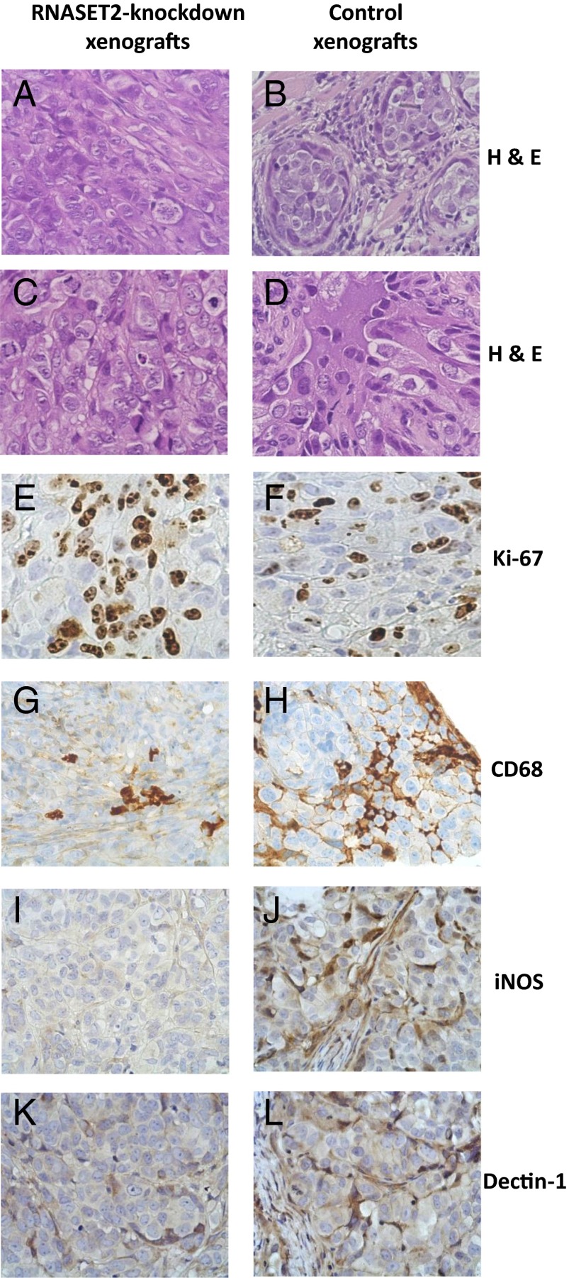
Fig. 2.
Histological and immunohistochemical analysis of tumor samples. Parental and RNASET2-silenced OVCAR3-derived tumor sections were stained with H&E (A–D). Microphotographs show a prevailing diffuse pattern and some solid nests in the central areas of RNASET2-silenced tumors, filled by large and polygonal cells with prominent nuclei and nucleoli (A and C). By contrast, control tumors show widespread nests/micronests pattern, with focal slender fibrous septa (B and D). Tumor sections were also analyzed by IHC for Ki-67 as a marker for cell proliferation (E and F) and for CD68 (G and H), inducible Nitric Oxide Synthase (iNOS-1) (I and J), and dectin-1 (K and L) to detect murine host inflammatory cells, the whole macrophage cell population, and M1- and M2-polarized macrophages, respectively. Photomicrographs shown are representative images of multiple fields examined in at least four sections derived from tumor excised in two to three independent animals for each experimental group.

