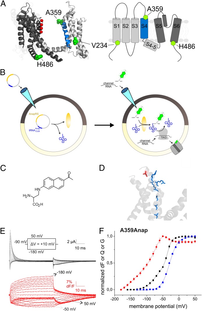
Fig. 1.
(A) Structure of KV1.2/2.1 chimera (PDB ID code 2R9R, two subunits) and topology (transmembrane segments S1–S6). The position of the mutations V234, A359, and H486 are displayed in green, and the potassium ions in the selectivity filter in red. (B) Principle of expression of fUAA. First, the plasmid to express the AnapRS and the corresponding tRNA is injected. On the subsequent day, fUAA and channel mRNA are injected, which leads to incorporation of the fUAA into the channel. (C) Structure of Anap. (D) Position of A359 (red) with respect to the arginines in the S4 (blue; PDB ID code 2A79). (E) Fluorescence response and gating currents of A359Anap in response to pulses from −90 mV to potentials between −180 and 50 mV. (F) Fluorescence voltage (FV, red circles), gating charge voltage (QV, black squares), and conductance voltage (GV, blue triangles) relations of A359Anap. The GV was fitted to a Boltzmann relation (V1/2 = −29.1 mV, dV = 12.6 mV). The QV and FV were fitted to a sum of two (V1/2,1 = −66.3 mV, dV1 = 19.1 mV, V1/2,2 = −35.2 mV, dV2 = 5.4 mV) and three Boltzmann relations (V1/2,1 = −134.6 mV, dV1 = 32.8 mV, V1/2,2 = −62.2mV, dV2 =15.1 mV, V1/2,3 = −41.6 mV, dV3 = 11.6 mV), respectively.

