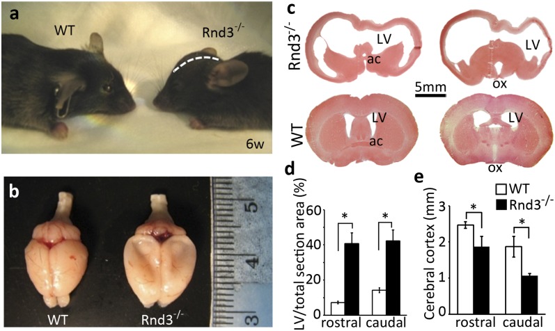
Fig. 2.
Rnd3-deficient mice developed hydrocephalus. (A) The mutant mouse displayed a domed skull (white dashed line) compared with the WT mouse. (B) A large amount of CSF was drained out during brain isolation, and the global brains showed the enlarged hemispheres, compressed olfactory bulbs, and subsided cerebral cortex in the mutant mice. (C) Representative brain coronal sections across the anterior commissure (ac) (Left) and optical chiasm (ox) (Right) showed the extreme dilations of the LVs along with a thin cerebral cortex in the mutant mice (Upper) compared with the WT mice (Lower). The brain sections were stained with H&E. (D and E) The area of the LV (D) and the thickness of the cerebral cortex (E) were quantified from a total of 12 staining pictures taken from six brains from 6-wk-old mice in each group. *P < 0.05 vs. WT.

