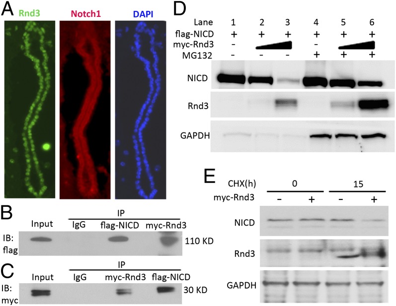
Fig. 5.
Rnd3 physically interacted with NICD and facilitated its degradation. (A) Coronal sections of a WT mouse aqueduct showed endogenous Rnd3 (green) and Notch1 (red) expression by immunofluorescent staining. (B and C) Co-immunoprecipitation (IP) pull-downs were conducted, followed by immunoblotting analyses (IB). The blots confirmed the interaction of Rnd3 and NICD proteins. (D) NICD protein levels in the entire Rnd3 expression vector transfected cell lysates were measured by immunoblot analysis. Along with the elevated Rnd3 protein levels, noticeable decreases in NICD protein levels were observed; this effect was attenuated by treatment with a proteasome inhibitor, MG132. (E) Immunoblot analysis showed that the degradation of endogenous NICD was enhanced by the introduction of Rnd3 when protein synthesis was blocked by treatment with cycloheximide (CHX). h, hours.

