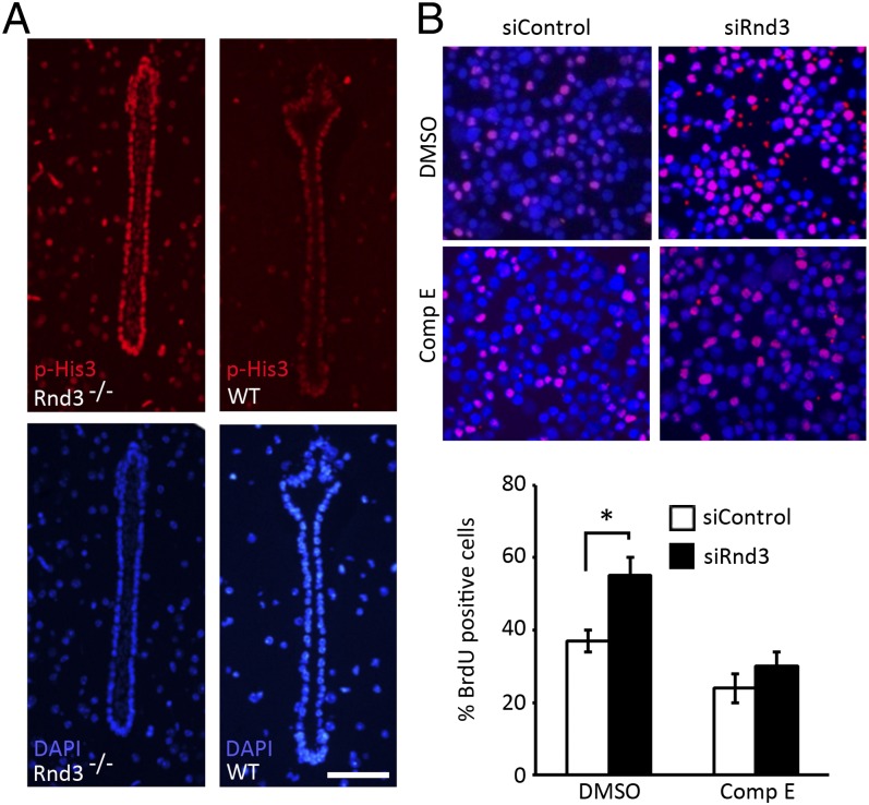
Fig. 6.
Genetic deletion of Rnd3 promoted aqueductal ependymal cell proliferation, which was attenuated by treatment with compound E (Comp E). (A) p-His3 was detected in Rnd3−/− aqueduct ependymal cells. The lower panels were DAPI staining. (B) A noticeable increase in BrdU-positive cells (pink) was observed in the HEK 293T cells treated with the siRNA specific for Rnd3 (siRnd3) (upper right section of the image). This increase was attenuated by treatment with compound E (lower right section of the image). The BrdU-positive cells were quantified from nine images, taken from three slides, and presented in the lower panel. (Scale bar: 25 μm.) *P < 0.05 vs. control siRNA (siControl) group.

