Fig. 1.
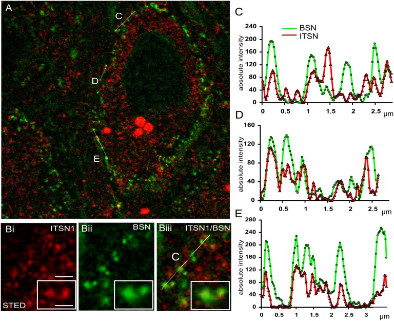
Intersectin 1 is localized at and around AZs. (A) STED microscopy image of a mature mouse calyx of Held synapse (age P50–P70) immunostained for endogenous bassoon (BSN; green) and intersectin 1 (ITSN1; red). The postsynaptic neuron is surrounded by the calyx terminal immunopositive for the AZ marker bassoon. Note that bassoon staining is also detectable at noncalycial terminals. (Inset) Confocal image of bassoon immunopositive puncta. For the scale bar, see the line scan profiles in C–E. (B) Magnified images of the data shown in A (from line C). (B, i) ITSN1; (B, ii) BSN; (B, iii) merged image. (Insets) Intersectin-immunopositive puncta confined to the active zones. (Scale bars: 750 nm; 350 nm in Insets.) (C–E) Spatial intensity profiles of bassoon (green) and intersectin 1 (red). The regions of interest (lines C, D, and E in A) were selected where the presynaptic compartment showed a finger-like structure.

