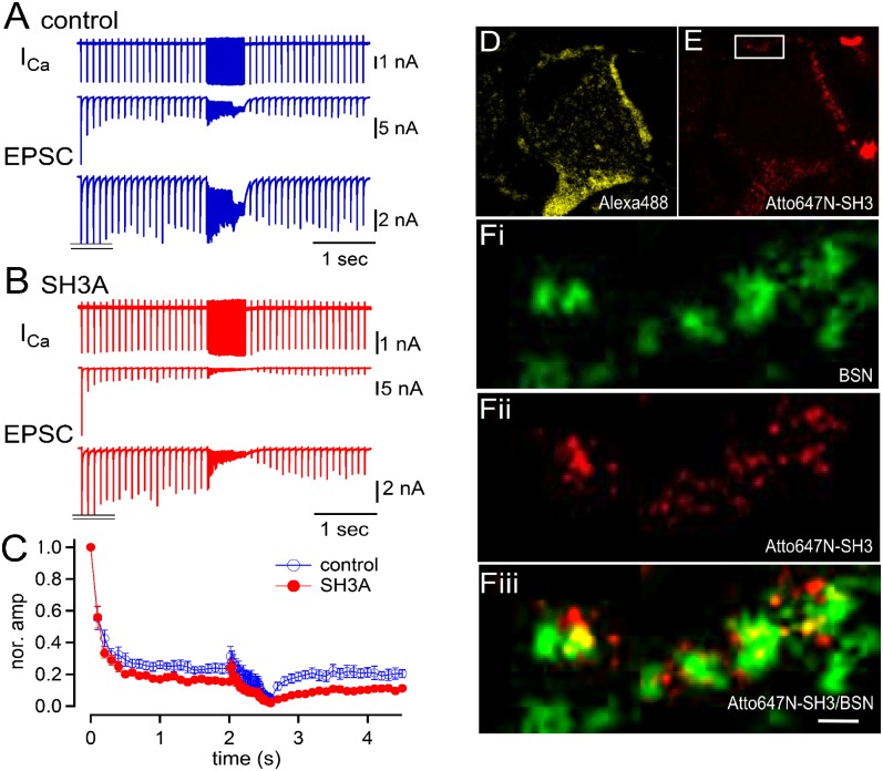
Fig. 3.
Acute perturbation of intersectin 1 SH3A domain function causes short-term synaptic depression. Simultaneous recordings of the presynaptic and postsynaptic compartments at the calyx of Held synapse (rats, P8–P11). A train of AP-like stimuli (depolarization to +40 mV for 1.5 ms) was applied (20 pulses), at 10, 50, and 100 Hz, and then at 10 Hz. Presynaptic Ca currents (Top), EPSCs (Middle), and magnified EPSCs (with initial EPSC peaks truncated; Bottom) are shown. (A and B) Data under control conditions (A) and in the presence of SH3A domain (5 μM) (B). (C) Normalized time course of the EPSC amplitudes plotted over time. Red and blue symbols indicate the data under control and in the presence of the SH3A domain, respectively. (D and E) Confocal images of a calyx terminal (rats, P8–P11) preloaded with Alexa Fluor 488 (200 μM; D) and the Atto647N-conjugated SH3A domain of intersectin (10 μM; E). (F) STED images of magnified data outlined by the box in E, illustrating the localization of the AZ markers bassoon (F, i; green), and Atto-SH3A (F, ii; red). (F, iii) Composite image, with colocalizing puncta in yellow. (Scale bar: 500 nm.)

