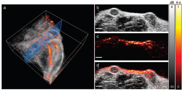
Fig. 5.

In vivo anatomical photoacoustic and ultrasound images. (a) 3D overlay of photoacoustic and ultrasound images of the upper leg/abdominal region. Blue box shows the 2D imaging plane of (b–d). (b) Ultrasound image showing two hypoechoic tumors. (c) Photoacoustic image, acquired using a laser wavelength of 850 nm. (d) Overlay of photoacoustic and ultrasound images. Scale bars = 2 mm.
