
Fig. 7.
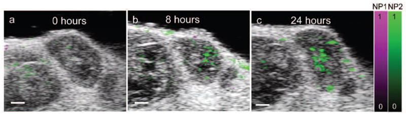
In vivo molecular imaging of contrast agent accumulation within a targeted tumor. The multiwavelength photoacoustic signal was unmixed into two components corresponding to two different contrast agents, nanoparticle 1 (NP1) and nanoparticle 2 (NP2). (a) 2D overlay of ultrasound image, distribution of NP1 and NP2 before the injection of the contrast agent, showing minimal background photoacoustic signal. Accumulation of NP1 and NP2 within the tumor is shown (b) 8 hours and (c) 24 hours after intravenous injection. Scale bars = 1 mm.