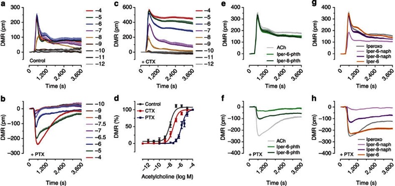
Figure 3. Cellular DMR reveals structure-dependent Gi bias of dualsteric probes.
(a–c) Shown are baseline-corrected DMR responses measured as wavelength shifts (pm) and recorded over time after addition of indicated concentrations of ACh in non-pretreated CHO-hM2 cells (control, a) and either PTX (b)- or CTX (c)-pretreated CHO-hM2 cells (means and s.e.m.) from one representative out of at least four independent experiments conducted in quadruplicates. (a) ACh exposure of control cells induces concentration-dependent positive DMR with peaks reflecting Gi signalling. (b) Pretreatment with the Gi inhibitor PTX (100 ng ml−1, 16–24 h) reveals negative DMR reflecting Gs signalling. (c) Masking Gs-dependent signalling by CTX pretreatment (100 ng ml−1, 8 h) induces positive DMR followed by sustained plateaus at higher concentrations. (d) Concentration-effect curves of ACh-induced peak DMR; data (means±s.e.m. from at least four experiments) are normalized to the response obtained with 100 μM ACh. The pronounced sensitivity of ACh's potency to PTX indicates Gi dominance in the control DMR peaks. (e–h) DMR recordings of indicated compounds in either control cells (e,g) or PTX-pretreated cells (f,h). Applied were maximally effective concentrations: ACh 100 μM, iper-6-phth 100 μM, iper-8-phth 10 μM, iperoxo 1 μM, iper-6-naph 100 μM, iper-8-naph 10 μM and iper-6 100 μM. Representative data (means and s.e.m.) of at least three independent experiments conducted in quadruplicates.

