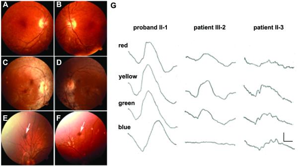
Figure 2.
Clinical data of the American family. A-F) Fundus photographs. The proband II-1 shows a few small spots of clumped pigmentation in her peripheral retina, mild atrophic changes of the retinal epithelium in the macula, but no attenuation of retinal vessels (A, B). Her sister II-3 shows attenuated retinal vessels and clumped intraretinal pigmentation in her mid-peripheral retina (C, D). Patients IV-1 (E) and IV-2 (F) show a retinal pigmentary clump (arrow) in the peripheral retina. G) ERGs of patients in the American family. The subjects were studied in the dark-adapted state using Wratten filters providing red (Wratten 29), yellow (Wratten 21), green (Wratten 61) and blue (Wratten 98) light (see Methods). The proband, her son and her sister were analyzed in 1980, when the two most severely affected individuals had detectable responses. The proband, who is the oldest of all affected, has much better responses than her sister II-3 and her son III-2. Patient II-3 response to blue light has greater amplitude than that of III-2 (arrows). The converse is true for red light (arrows). The calibration, lower right, indicates 7 microvolts vertically and 20 milliseconds horizontally.

