Figure 6.
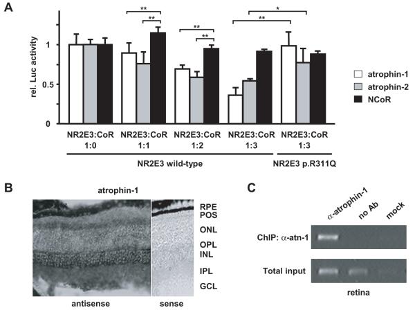
Atrophins are corepressors of NR2E3. a) Atrophins enhanced NR2E3-mediated repression of the M-opsin promoter in presence of NR2E3 wild-type, but not NR2E3 p.R311Q protein. 293T cells were transiently transfected with the M-opsin reporter construct, in presence of NRL, CRX, and, respectively, NR2E3 wild-type or p.R311Q (30 ng each), along with increasing amounts of atrophin-1, atrophin-2 and NCoR (33 ng, 66 ng or 100 ng). At least three independent experiments were performed in duplicates in 12-well plates using the luciferase assay system (Promega). Luciferase activity was normalized to β-galactosidase activity. Normalized luciferase activities of cells transfected without corepressors were set to 1. Error bars represent S.E.M; *: p<0.05; **: p<0.01. B) In situ hybridization for atrophin-1 mRNAs on retinal sections of 2-months-old Bl6/C57. RPE: retinal pigment epithelium; POS: photoreceptor outer segments; ONL: outer nuclear layer; OPL: outer plexiform layer; INL: inner nuclear layer; IPL: inner plexiform layer; GCL: ganglion cell layer. Note the absence of signal in presence of the control sense probe. Scale bar: 50 μm. C) Atrophin-1 associates with the M-opsin promoter in vivo. Chromatin-immunoprecipitation for the M-opsin promoter was performed with anti-atrophin-1 antisera (α-atn-1) on retinas of 2-months-old C57/BL6 mice. As controls, immunoprecipitation without antisera (no Ab) or with the buffer alone (mock) were used. PCR amplifications on total input chromatin (Total input) are shown in the lower panel.

