Figure 4.
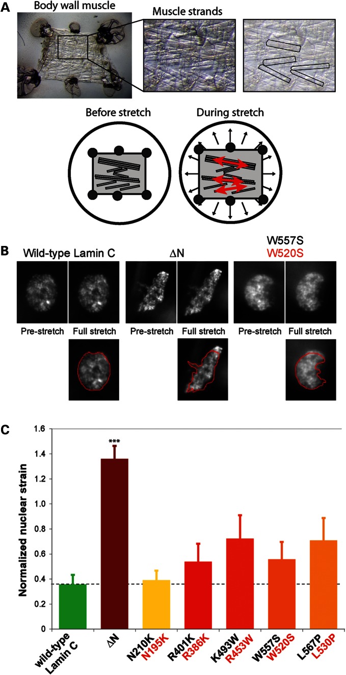
Decreased nuclear stiffness in body wall muscle of D. melanogaster larvae expressing Lamin C variants causing EDMD. (A) Overview of the experimental approach to observe nuclear deformations in D. melanogaster larval filet subjected to strain application. Upper panel: the larval filet, consisting of the body wall muscle, is glued to a transparent silicone membrane (left panel); individual muscle fibers are easily detectable (middle and right panel, with distinct muscle fibers highlighted in the right panel). Lower panel: although biaxial strain is applied to the Drosophila larval filet, the tissue strain in the muscle fibers is almost completely uniaxial in the direction of the muscle fiber. (B) Change in nuclear shape in pre-stretched and fully stretched muscle strands of larvae expressing wild-type Lamin C (the A-type lamin of D. melanogaster), head-truncated ΔN Lamin C or the W557S Lamin C mutation, which corresponds to the human LMNA W520S mutation. Muscle cells expressing the ΔN lamin mutation deformed significantly more under applied strain. Images in the lower panel represent nuclei under strain, overlaid with the nuclear contour from the pre-stretch state in red. (C) Normalized nuclear stiffness of D. melanogaster larvae expressing wild-type D. melanogaster Lamin C, head-truncated Lamin C ΔN or Lamin C mutations (labeled in black) that correspond to human EDMD-causing LMNA mutations (labeled in red). ***P < 0.001, compared with muscle nuclei expressing wild-type Lamin C.

