Figure 5.
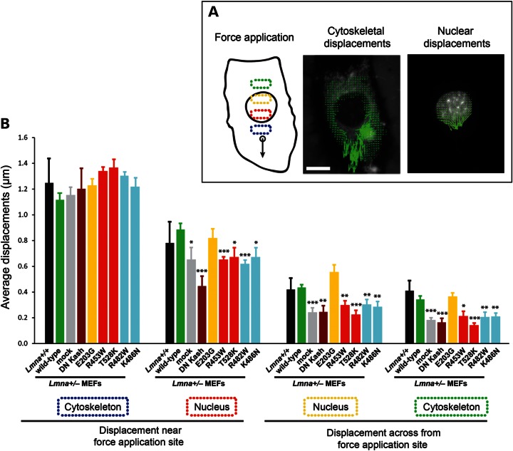
Laminopathic lamin A mutations disrupt force transmission between the nucleus and cytoskeleton. (A) Schematic overview of the microneedle manipulation experiments to assess intracellular force transmission between the nucleus and cytoskeleton. A fine microneedle is inserted into the cytoplasm near the nucleus and moved toward the periphery (left panel). Displacement maps of the cytoskeleton (middle panel) and the nucleus (right panel) are plotted by tracking intracellular fluorescent markers; measurements of the average displacements within four defined areas inside the cytoplasm and nucleus (indicated by dotted boxes in the left panel) are used to evaluate the intracellular force transmission between the nucleus and cytoskeleton. Scale bar: 20 µm. (B) Average displacements at the indicated regions in Lmna+/+ MEFs, and in Lmna+/− fibroblasts stably expressing the empty vector (mock), wild-type lamin A, head-truncated ΔNLA or disease-specific lamin A mutations. The displacements in the first cytoskeletal region were comparable in all cells, illustrating similar strain application with the microneedle. Lmna+/− cells expressing mutant lamins (except for the E203G mutations) and the empty vector had significantly smaller nuclear and cytoskeletal displacements in the three cellular regions observed away from the site of strain application compared with Lmna+/− cells expressing wild-type lamin A or non-modified Lmna+/+ cells, indicating reduced intracellular force transmission between the nucleus and cytoskeleton in the mutant cells. *P < 0.05; **P < 0.01; ***P < 0.001 versus Lmna+/− MEFs expressing wild-type lamin A.

