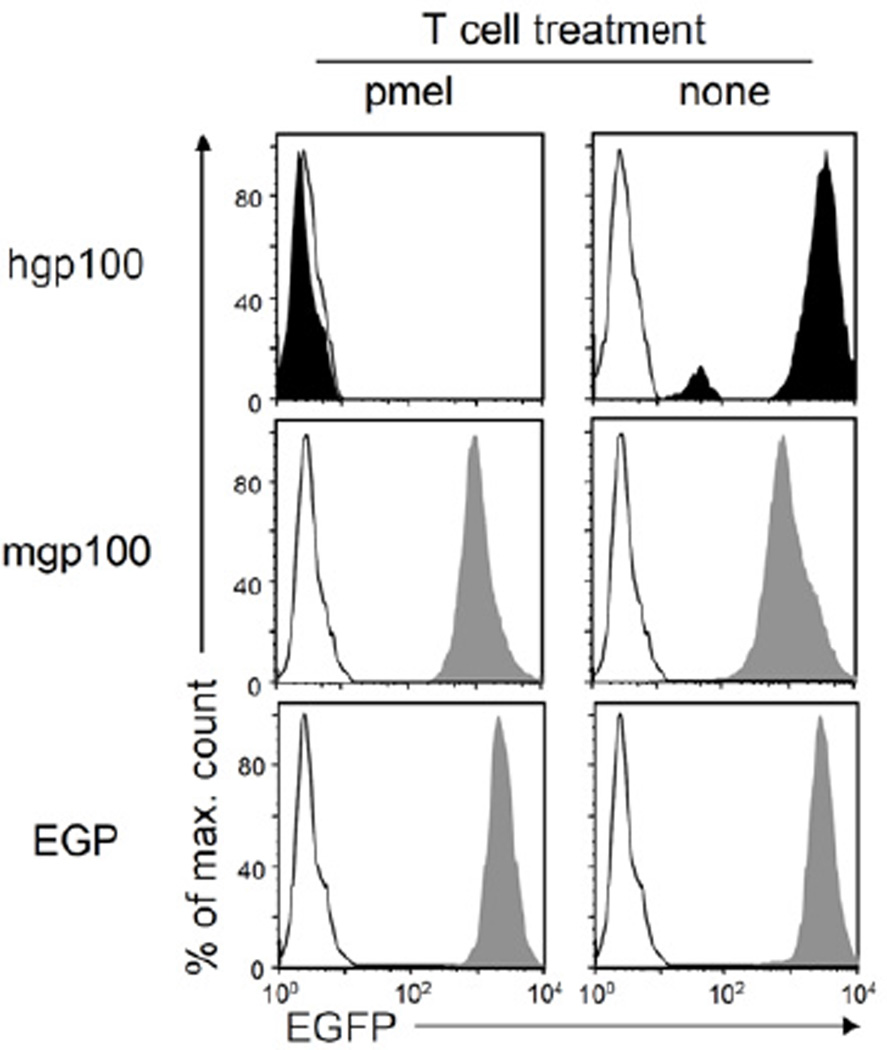
Figure 3. Outgrowth of antigen-loss variants after pmel T cell treatment of cancer cells expressing hgp10025 but not of cancers expressing mgp10025 or EGP.
Cancer cells of relapsed tumors expressing mgp10025, EGP (both gray) or hgp10025 (black) were isolated after pmel T cell treatment, adapted to culture, and analyzed for peptide-EGFP fusion gene expression (left panels). MC57 cells (white histogram) cultured in vitro and MC57-mgp100, MC57-EGP (both gray) or MC57-hgp100 (black) cells isolated from non-treated mice (right panels) were analyzed as controls. Isolated lines from mgp100- or hgp100-expressing tumors are representative for four lines each, the isolate from the EGP-expressing tumor is representative for two lines; all lines were isolated after relapse (respective tumors were marked with * in Figure 2B). The repeatability in independent experiments strongly suggests that loss of antigen expression from the hgp10025 cancer cells was not an artifact caused by adaptation or post-isolation culturing. See also Figure S3.

