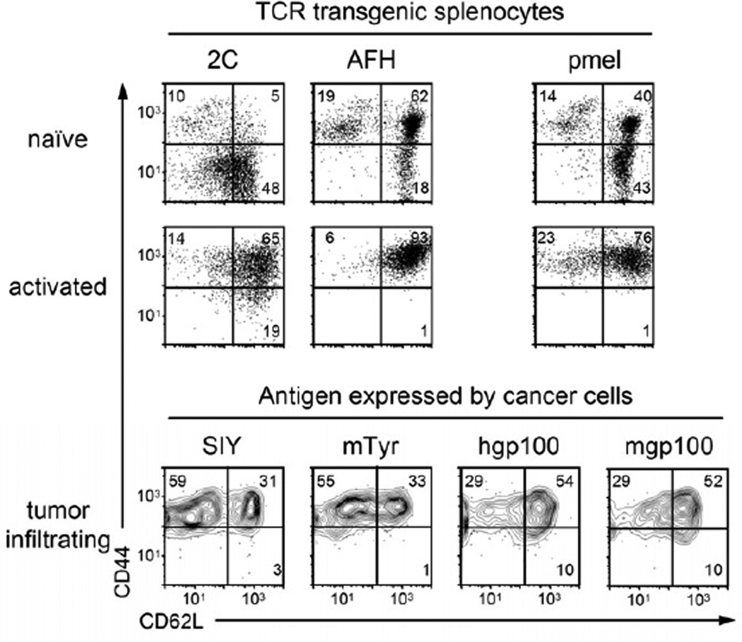
Figure 4. T cells transferred to treat the tumors expressing the different peptides showed the same phenotype of activated T cells.
T cells of 2C AFH and pmel TCR-transgenic mice were tested for their activation status in “naïve”, untreated mice (splenocytes), at day 3 after peptide activation in vitro, and on day 4 after adoptive transfer (tumor infiltrating cells). Cells were analyzed by flow cytometry for expression of CD44 and CD62L, and gated on CD8+ T cells expressing the cognate Vβ-chain: Vβ8 for 2C, Vβ11 for AFH and Vβ13 for pmel. Data are representative for two or three independent experiments for the data in vivo and in vitro data, respectively.

