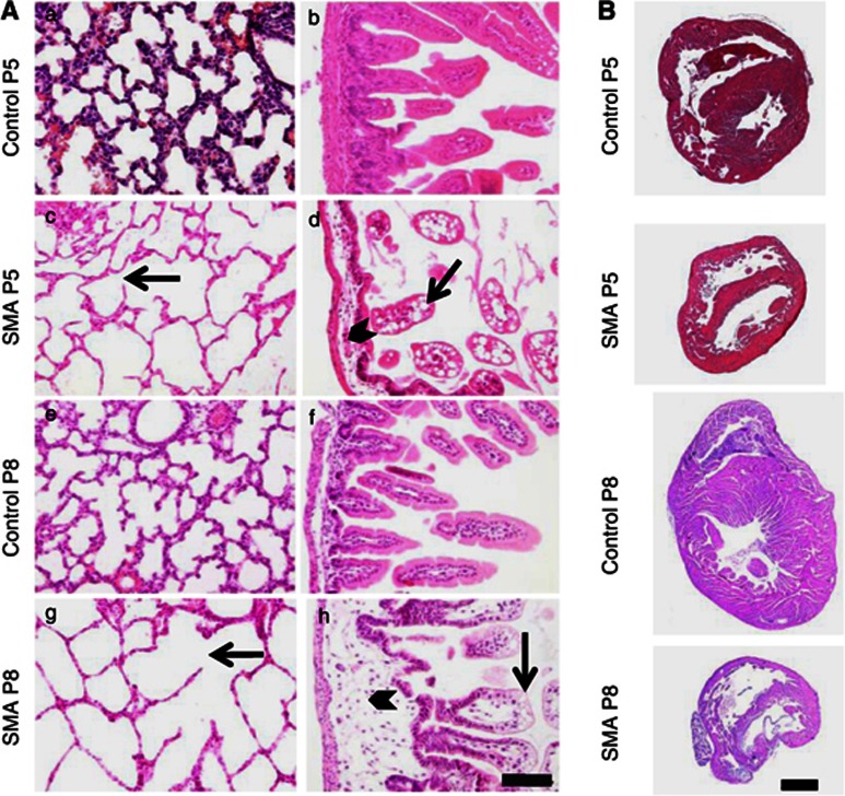
Figure 4.
Histology of the lung, intestine and heart of SMA animals compared with control littermates at P5 and P8. (A) Mice at P5 and P8 reveal similar lesions at different degrees of severity. SMA animals (c, g) show variable degrees of emphysema with ruptured alveolar septa and enlarged alveolar spaces compared with control animals (a, e). The small intestine reveals reduced numbers of villi, and villi are blunt and club-shaped with severe intramural edema in the lamina propria of SMA animals (arrowheads in d, h). Mucosal epithelial cells show intracytoplasmatic vacuoles predominantly at the tips of the villi and lacteals were occasionally dilated (arrows in d, h). These lesions are absent in age-matched controls (b, f). Changes were obvious at P5 and show progression towards P8. Scale bar 100 μm. (B) Representative pictures of hearts of control and SMA animals at P5 and P8. Interventricular septa appear thinned in SMA animals.

