Figure 3.
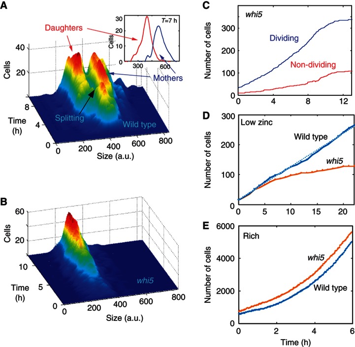
No population splitting in whi5. (A) Mothers and daughters differ in cell size. The distributions of cell sizes of wild-type mothers and daughters at different times following the transfer to LZM. The inset shows the distribution at t=7 h after accounting for age differences. (To make sure the size difference is not owing to the fact that mothers are older than the daughters, we treated the single-cell growth curves as if all cells were born at the same time.) (B) Deletion of WHI5 eliminates the splitting into subpopulations. Same as (A), for whi5 cells. (C) Deletion of WHI5 enables division of daughter cells. Increase in the number of dividing (blue) and non-dividing (red) cells in multiple colonies of whi5 cells. Note that in contrast to the wild-type where the number of dividing cells (Figure 2b, blue) stops increasing about 2 h after the shift, in whi5 the number of dividing cells continues to grow long time after the shift. Thus, even daughter cells that were born in low zinc divide. (D, E) Proliferation of wild-type and whi5 cells. Growth curves of wild-type and whi5 cells grown together in the same flow cell in low zinc (D) and rich (E) media. See also Supplementary Figure 3.

