Figure 7.
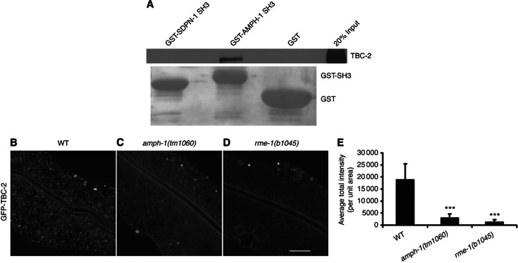
Experimental validation of predicted interactions and endocytosis functions in worm. (A) The worm AMPH-1 SH3 domain physically interacts with TBC-2 and not the SDPN-1 SH3 domain or GST controls, as shown by western blot. GST bait proteins, visualized by Ponceau S staining, are shown under the corresponding western blot lanes. (B–D) GFP-TBC-2 localization to endosomes (B) is disrupted in amph-1 (C) and rme-1 (D) mutants. Comparable confocal images show living intestinal epithelial cells expressing GFP-tagged TBC-2 from an integrated low-copy number transgene in wild-type, amph-1(tm1060), and rme-1(b1045) mutant animals. Scale bar, 10 μm. (E) Average fluorescence intensity of GFP-TBC-2 labeled puncta quantifies disruption of TBC-2 endosome localization across multiple experiments. Error bars, standard deviation (n=18 each, six animals of each genotype sampled in three different regions of each intestine). Significant differences in the one-tailed Student’s t-test are indicated (***P=0.001).

