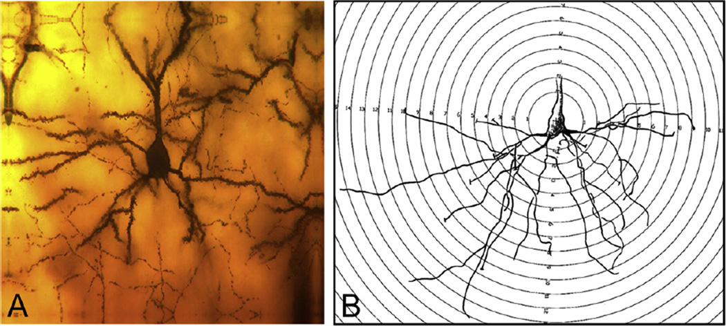
Fig. 6.
The extent and distribution of neuronal dendritic branching in the parietal cortex, layers II–III, were evaluated by Sholl analysis. (A) Typical parietal neuron from our study. (B) Transparent overlay of increasingly larger concentric circles at 10 lm intervals was superimposed on the camera lucida drawings. The number of dendritic branch intersections with each progressively larger circle is counted from the soma of each neuron. This generates a profile showing the amount of dendritic branching material.

