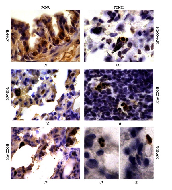
Figure 5.

(a–c) Cell proliferation and (d–g) apoptosis, detected by PCNA immunolabelling and TUNEL staining, respectively, after in vivo exposure to 1 mg/kg b.w.: (c, d, e) MW-COOH and (a, b, f, g) MW-NH2. PCNA-positive epithelial cells at (a) bronchiolar and (b, c) alveolar levels; TUNEL-positive CNT-laden macrophages in both (d) normal and (e) inflammatory stromal areas as well as (f, g) at alveolar levels. Objective magnification: 60x (a–e), 100x (f, g).
