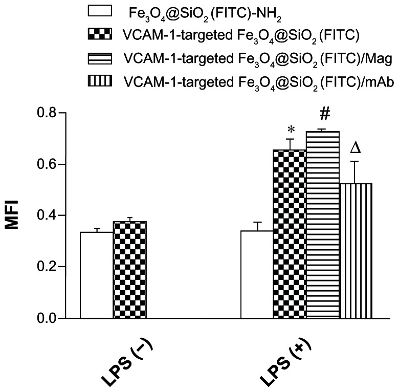
Figure 7.
Adhesion of fluorescent nanoparticles on a monolayer of HUVEC-CS under static conditions calculated quantitatively using MFI.
Notes: The nanoparticles were cocultured with HUVEC-CS for 45 minutes, and washed three times with phosphate-buffered saline to remove the nonadhered nanoparticles. The adhered nanoparticles were observed under an inverted fluorescence microscope. *P < 0.05 versus Fe3O4@SiO2(FITC)-NH2 at LPS (+); #P < 0.05 versus VCAM-1-targeted Fe3O4@SiO2(FITC) at LPS (+); ΔP < 0.05 versus VCAM-1-targeted Fe3O4@SiO2(FITC) at LPS (+) lipopolysaccharide.
Abbreviations: Fe3O4@SiO2(FITC), fluorescein isothiocyanate-loaded silica-coated superparamagnetic iron oxide nanoparticles; VCAM-1, vascular cell adhesion molecule-1; MFI, mean fluorescence intensity; LPS, lipopolysaccharide; mAb, monoclonal antibody; LPS (−): no LPS stimulation; LPS (+): stimulation with 1 μg/mL LPS for 5 hours; LPS (+)/Mag: LPS stimulation for 5 hours plus magnetic field; LPS (+)/monoclonal antibody: LPS stimulation for 5 hours plus anti-VCAM-1 monoclonal antibody pretreatment; HUVEC-CS, an inflammatory subline of human umbilical vein endothelial cells.

