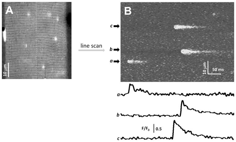
Fig. 2.

(a) A xy field scan image of a permeabilized EDL muscle fiber loaded with the internal solution containing 100 μM fluo-4, 200 nM free Ca2+, and 2 mM free Mg2+. (b) A xt line scan image of the same muscle fiber obtained through repeatedly scanning the fiber along the dashed line. Traces a, b, and c are the quantitative measurement of Ca2+ sparks by evalu-2+ sparks.
