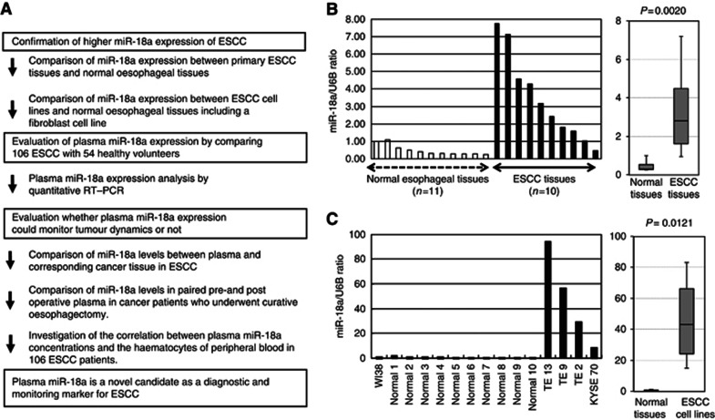
Figure 1.
(A) Study design to develop a novel biomarker of plasma microRNA. (B) The expression of miR-18a in ESCC tissues. Differential expressions of miR-18a in ESCC tissues were compared with those of normal tissues by a waterfall plot (A). Expression levels of miR-18a were significantly higher in ESCC tissues than normal oesophageal tissues (P=0.0020). The upper and lower limits of the boxes and lines inside the boxes indicate the 75th and 25th percentiles and the median, respectively. Upper and lower horizontal bars denote the 90th and 10th percentiles. (C) The expression of miR-18a in ESCC cell lines. Differential expressions of miR-18a in ESCC cell lines were compared with those of a fibroblast cell line and normal tissues. In ESCC cell lines, expression levels of miR-18a were significantly higher than in both a fibroblast cell line and normal oesophageal tissues (P=0.0121). *Normal means normal tissues including the normal fibroblast cell line, such as WI-38.

