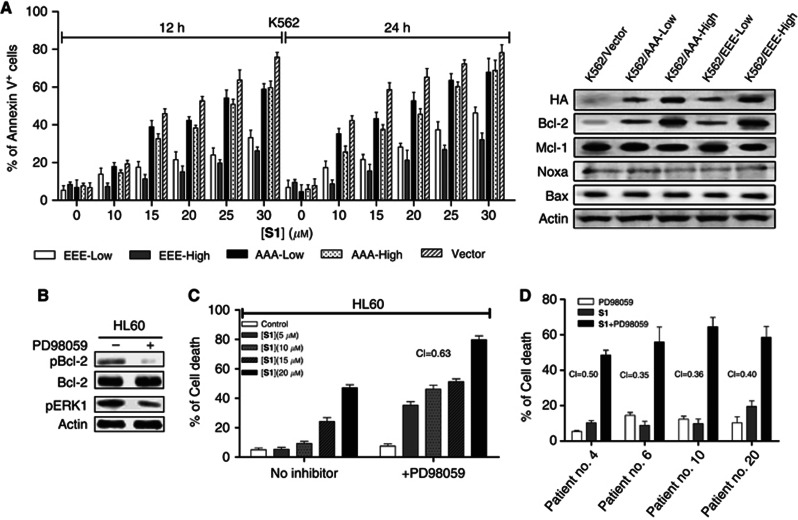
Figure 3.
Bcl-2 phosphorylation modulates sensitivity to S1 in leukaemic cells. (A) K562-Vector cells and K562 expressing different levels of mutant Bcl-2 cells were treated with S1 for 12 or 24 h and Annexin V+ cells were analysed. Indicated protein levels were examined by immunoblotting. Apoptosis was quantified by Annexin V staining and determined by flow cytometric analysis. Results represented the mean of three independent experiments. (B) HL60 cells were treated for 24 h with 20 μℳ PD98059 and total protein extracts were analysed by western blot for the expression of pERK1, pBcl-2(Ser70), Bcl-2 and actin. (C) HL60 cells were treated with the indicated doses of S1 alone or together with 20 μℳ PD98059 for 24 h. Viability was assessed by 3-(4,5-dimethythiazol-2-yl)-2,5-diphenyl tetrazolium bromide (MTT) assay. Combination indexes (CIs) were calculated using the Calcusyn software and indicated inside the plot for each combination. (D) Cells from four resistant leukaemia patients were cotreated with 20 μℳ PD98059 and 20 μℳ S1 for 40 h and viability was assessed by MTT assay. Combination indexes were calculated as above.

