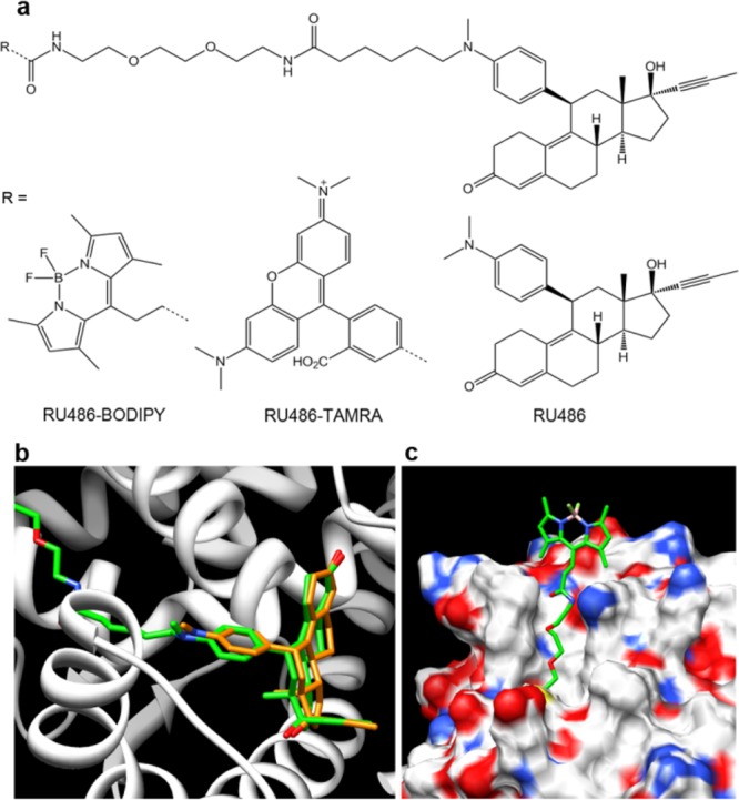
Figure 1.

Fluorescent ligands of progesterone receptor. (a) Chemical structures of RU486 and its fluorescently labeled derivatives RU486-BODIPY and RU486-TAMRA. Molecular modeling of RU486-BODIPY (green) bound to human PR showing (b) orientation inside the ligand binding pocket compared to RU486 (orange) (amino acids 712–720 have been removed for clarity) and (c) the linker extends out of the binding pocket, placing the BODIPY dye well outside the protein shell.
