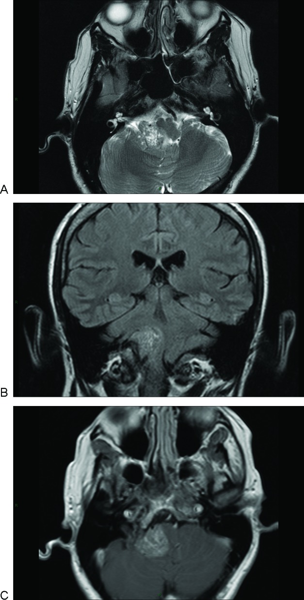
Figure 1.

Magnetic resonance imaging scan of primary cerebellopontine angle melanocytoma. (A) Axial T2 image showing a right cerebellopontine angle (CPA) isointense mass and areas of hypo and hyper intensity. (B) Coronal T1 image showing a right CPA mass displacing the brainstem. The lesion was slightly hyperintense with areas of isointensity. (C) Axial T1 with contrast demonstrating mild homogeneous enhancement.
