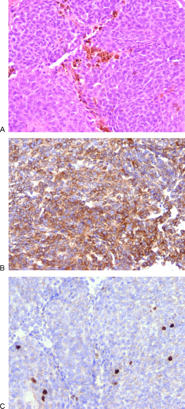
Figure 2.

Histological features of melanocytoma. All original magnifications are x200. (A) Hematoxylin and eosin stain demonstrating solid lobules of uniform cells with scanty pigment (mostly in macrophages) and rounded nuclei. (B) Immunocytochemistry for Melan-A showing strong cytoplasmic positivity, confirming the melanocytic nature and helping to exclude meningioma and schwannoma. (C) Immunocytochemistry for Ki-67 revealing a low proliferation index (~ 5%) which would be unusual in a primary or metastatic malignant melanoma.
