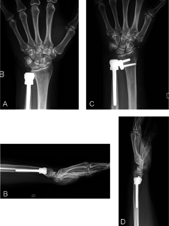
Figure 2.

(A and B) Preoperative PA and lateral radiograph of the wrist of Patient 3. The patient had undergone a previous ulnar head replacement for (A) primary distal radioulnar joint arthritis. Note erosion of the ulnar head into the sigmoid notch with overlying shelf of bone, which was felt to limit pronation. (C and D) Final postoperative PA and lateral radiographs showing a well-seated implant with no evidence of dorsal subluxation. Tapering of the ulnar shaft can be seen in the PA radiograph. PA, posterior to anterior.
