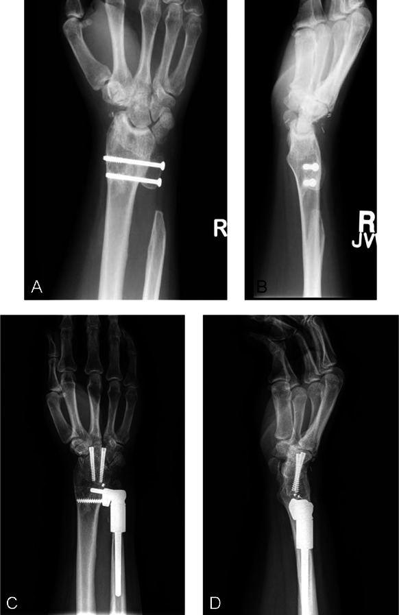
Figure 3.

(A and B) Preoperative PA and lateral radiograph of patient 4. The patient had undergone a previous radioscapholunate fusion and Sauvé–Kapandji procedure for wrist pain. At the time of initial evaluation, she had painful impingement of the proximal ulnar stump. (C and D) Final follow-up radiographs showing a well-seated implant. An extended collar was used to allow the sigmoid notch to be placed in position of the previously fused ulnar head. Note tapering of ulnar shaft on the PA radiograph. Lateral radiographs show good alignment. PA, posterior to anterior.
