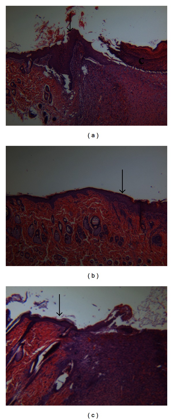
Figure 3.

Histological staining of sections from rat wounds at day 7. (a) H&E staining of untreated wound; (b) H&E staining of wound treated with CMHA-S gel; (c) H&E staining of wound treated with CMHA-S film. Arrowheads indicate the interface between native rat skin and neos-skin; c = crust; H&E staining, 50x.
