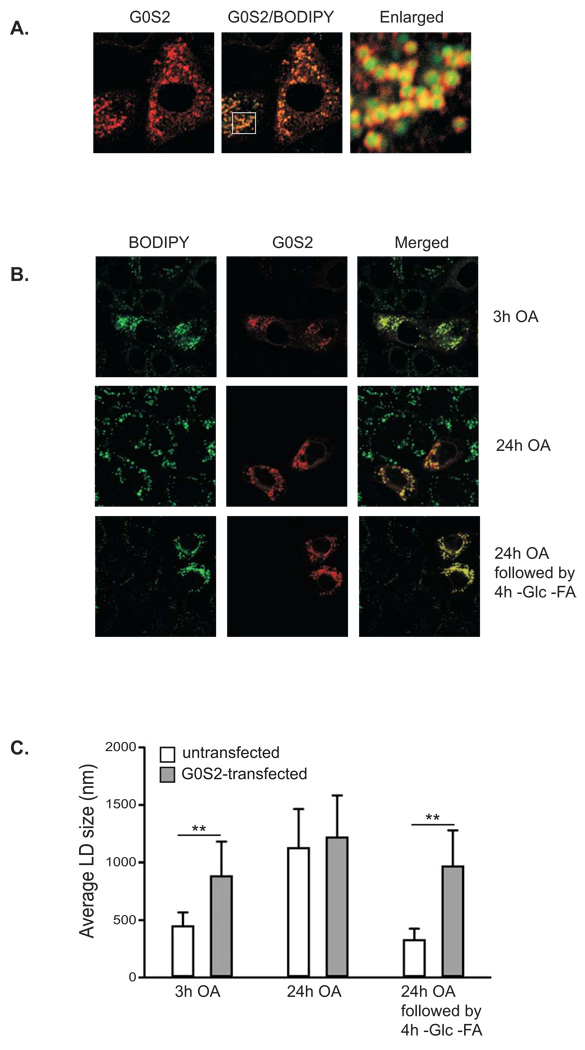
Figure 2. Overexpression of G0S2 inhibits lipid droplet degradation mediated by nutrient withdrawal.
A. Immunofluorescence staining with anti-G0S2 antibodies was performed to reveal localization of overexpressed G0S2 in HeLa cells. Lipid droplets were co-stained with BODIPY 493/503 fluorescence dye. B. HeLa cells transiently expressing G0S2 were incubated under normal growth conditions with 400 µM of oleic acid complexed to albumin at a molar ratio of 8:1 for 3 h (upper panel) or 24 h (middle panel). A separate set of cells were incubated in serum-free and glucose-free medium for 4 h following 24 h of incubation with 400 µM of oleic acid (lower panel). Immunofluorescence staining with anti-G0S2 antibodies was performed to reveal the transfected cells. Lipid droplets were co-stained with BODIPY 493/503 fluorescence dye. C. Quantification of lipid droplet diameter. An average of 80 lipid droplets was measured for each point. Data are shown as mean ± SD, **p < 0.01, t-test.

