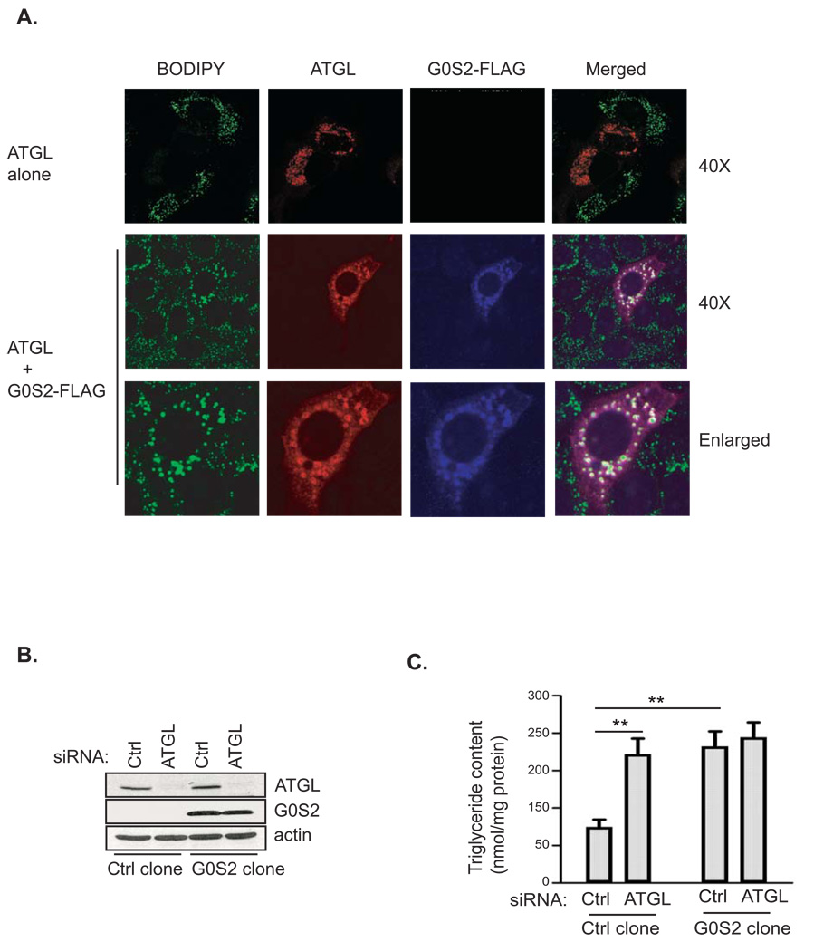
Figure 3. G0S2 inhibits lipid droplet degradation mediated by ATGL.
A. HeLa cells transfected with ATGL in the absence (upper panel) or presence (lower panel) of G0S2-FLAG were incubated under normal growth conditions with 400 µM of oleic acid complexed to albumin for 3 h. Immunofluorescence staining was performed by using anti-ATGL (red) and anti-FLAG (blue) antibodies. Lipid droplets were co-stained with BODIPY 493/503 fluorescence dye. B & C. Stable HeLa cell clones with or without untagged G0S2 expression were treated with ATGL siRNA (ATGL KD) or control mismatch siRNA (Ctrl KD). Protein expression was analyzed by immunoblotting with anti-ATGL and anti-G0S2 antibodies (B). The cells were incubated in 400 µM of oleic acid for 3 h and the intracellular TAG content was determined as described in Materials and Methods. The data were normalized with the total protein amounts in the cell extracts (C) (data are shown as mean ± SD and represent three independent experiments, **p < 0.01, t-test).

