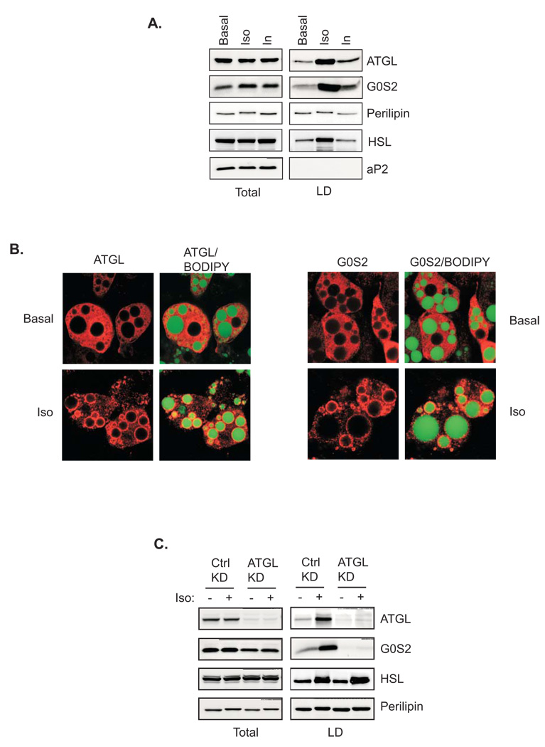
Figure 6. Lipid droplet localization of ATGL and G0S2 in adipocytes.
A. 3T3 L1 adipocytes were treated with or without 1µm isoproterenol or insulin for 30 min. Lipid droplets were isolated by ultracentrifugation. Total and lipid droplet-associated proteins were subjected to immunoblotting using antibodies against ATGL, perilipin, HSL, G0S2 and aP2. B. Immunofluorescence staining with anti-ATGL antibodies was performed to reveal localization of endogenous ATGL in 3T3-L1 adipocytes pretreated with or without 1 µM isoproterenol/0.25 mM IBMX for 30 min. Lipid droplets were co-stained with BODIPY 493/503. C. siRNA-mediated knockdown was performed by electroporating 3T3-L1 adipocytes with either control siRNA (Ctrl) or ATGL-specific siRNA (KD). 3 days later, cells were treated with or without 1 µM isoproterenol for 30 min followed by lipid droplets isolation. Total and lipid droplet-associated levels of ATGL, HSL, G0S2 and perilipin were analyzed by immunoblotting.

