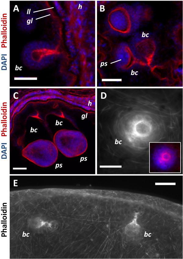
Figure 1.
Muscle fibers in the germinal layer and during brood capsule formation. All specimens stained with Phalloidin. A. Early brood capsule development (bc, brood capsule; gl, germinal layer; h, host tissue; ll, laminated layer). B. Brood capsules containing protoscolex buds (ps). C. Brood capsules during late protoscolex development. D. Whole-mount preparation showing an early brood capsule seen from above (inset shows merge with DAPI staining). Note the organization of the muscle fibers in the brood capsule region. E. General arrangement of the muscle fibers in the metacestode and brood capsules. A, B and C are sections from in vivo cultured material; D and E are whole-mount preparations of in vitro cultured material. Bars represent 50 μm.

