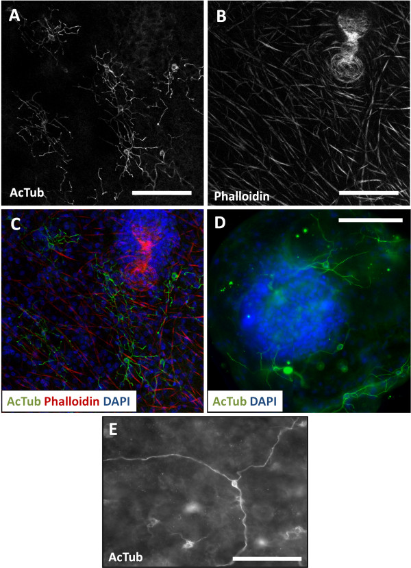
Figure 2.
AcTub-IR in whole-mount preparations of the germinal layer. A. AcTub-IR nerve cells in the germinal layer (in vitro cultured cyst, analyzed by confocal microscopy). B. Corresponding image of Phalloidin staining. C. Merge of AcTub-IR and Phalloidin staining combined to DAPI staining. Notice the lack of AcTub-IR cell bodies in the brood capsule. D. AcTub-IR nerve cells in a very small vesicle obtained from in vivo culture, containing only one brood capsule with a protoscolex bud (this image is a mosaic of three pictures and an overlay of several focal plains of epifluorescence microscopy). E. Unusual AcTub-IR nerve cell with long, unbranched neurites (in vitro cultured material). Neurites from other nerve cells are outside of the focal plane. Bars represent 50 μm.

