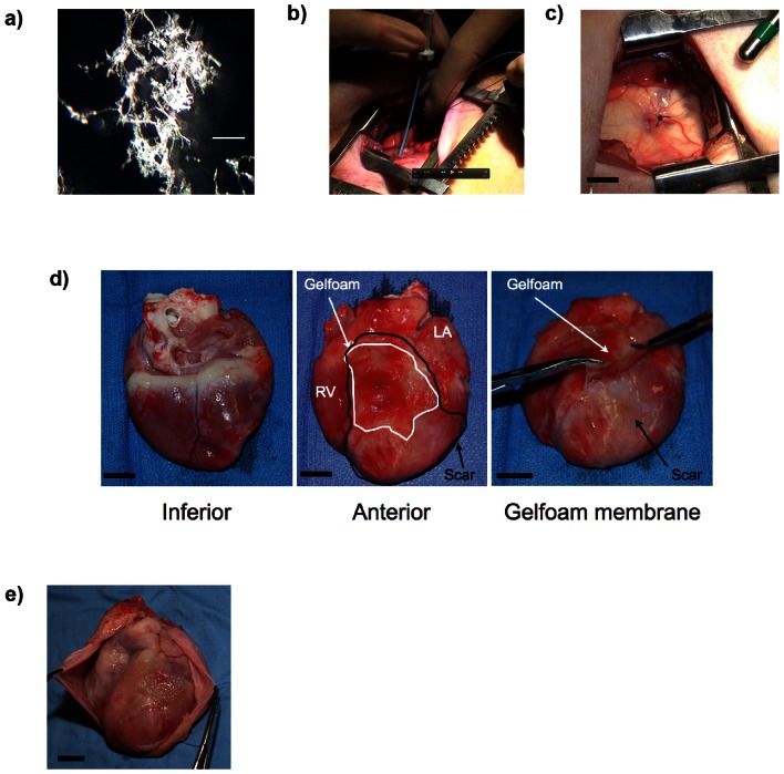
Figure 1. Pericardial application of periostin peptide using injectable gelfoam.
(a) Characterisation of homogenised gelfoam for injection. Scale bar 0.1 mm (b) Accessing the pericardial space with minimally invasive thoracotomy approach and injection of delivery system through an introducer. (c) View of the pericardium after injection of gelfoam. The delivery system appears white through the translucent pericardium. A purse string suture was placed to close the pericardial puncture site. (d) After 1 week, gelfoam (pink, outlined with black line) is located over the myocardial infarct scar (white, outlined with white line), while gelfoam is not located at the inferior surface. After 1 week, gelfoam has formed an elastic membrane, which can be peeled off. (e) Only minimal adhesions 3 months after application. Scale bars, 1 cm; RV, right ventricle.

