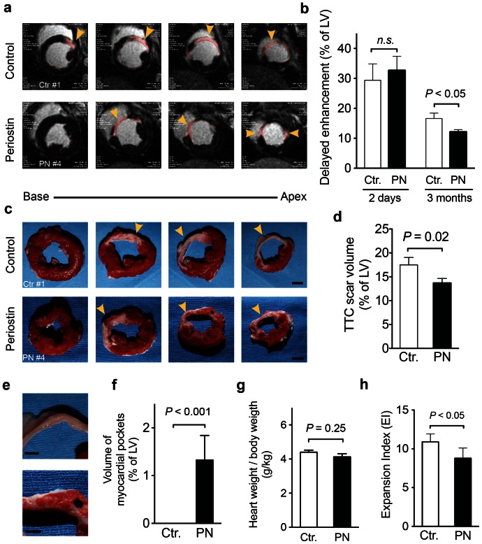
Figure 5. Periostin peptide-treated animals have smaller infarct scars and have myocardial strips.
(a, b) Quantification of scar volume by delayed enhancement (DE) MRI imaging 12 weeks after implantation of periostin peptide or control gelfoam. Representative examples showing DE in pink (a) and quantification as percentage of LV myocardium show smaller scar areas in periostin peptide-treated animals (b). (c, d) Quantification of scar volume by tetrazolium chloride (TTC) staining. Representative slabs of the left ventricle (c) and quantification (d) show smaller scar areas in periostin peptide-treated animals; Scale bars, 1 cm. Myocardial tissue was only detectable in the scar area of treated animals (e, f). The heart to body weight ratio remained unaffected, (g), while the expansion index was increased in swine receiving periostin (h). Statistical significance tested with t-test (b, d). Scale bars, 5 mm (e), 1 cm (c). PN (n = 7); Ctr (n = 6).

