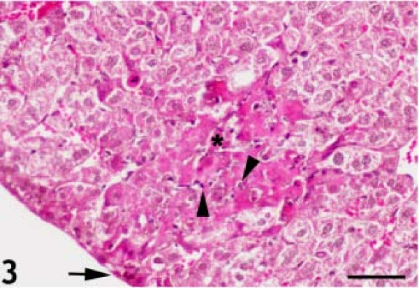
Figure 3.
Karyorrhexis and karyolysis in the nuclei of necrotic hepatocytes (arrowheads). There were several small inflammatory lesions on the surface of the liver (arrow); the number of lesions increased and progressed into deeper areas (asterisk). Hematoxylin and eosin staining. Bar: 50 micrometer.

