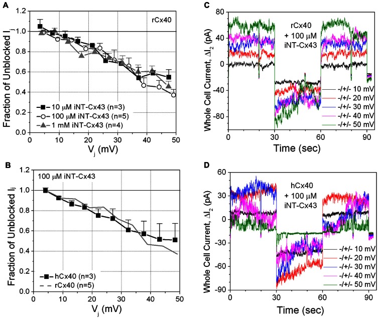
FIGURE 2.
(A) The inhibition of rCx40 gap junction currents (Ij) by unilateral addition of 10 μM (■), 100 μM (О), or 1.0 mM (∆) iNT-Cx43 peptide increased in a transjunctional voltage (Vj)-dependent manner. There was no statistical difference between the three curves based on comparison of the means at each Vj value. (B) The Vj-dependent blockade of hCx40 gap junctions (■) by 100 μM iNT-Cx43 peptide was not significantly different from rCx40 (continuous gray line). (C) Whole cell current traces recorded from cell 2 of a rat Cx40 cell pair with 100 μM iNT-Cx43 peptide added to cell 1 during the ΔV1 cation block voltage clamp protocol diagrammed in Figure 1B. The ΔI2 current (= -Ij) decrease during the +V1 voltage steps (only 10 mV incremental steps shown) illustrates the block induced by the iNT-Cx43 peptide. (D) Actual ΔI2 current recordings from a hCx40 cell pair during an iNT-Cx43 peptide experiment illustrating a similar Vj-dependent block of hCx40 gap junction currents. Gap junction channel currents are visible at ±50 mV with reduced open probability during the positive (blocking) Vj step.

