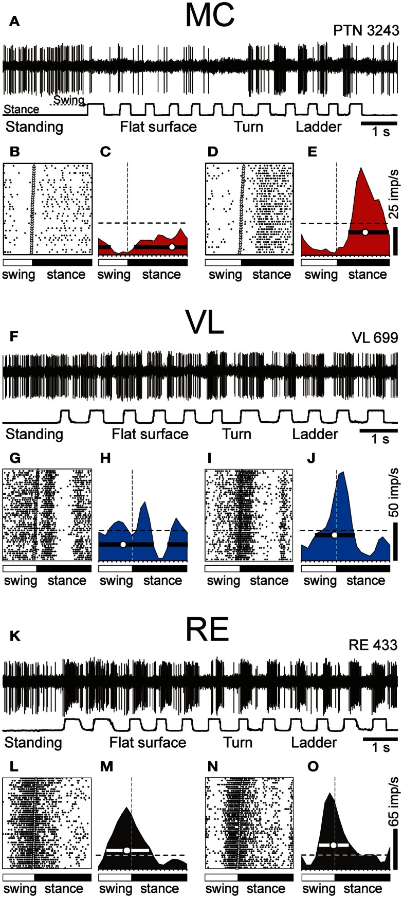
Figure 7.
Example activity of MC, VL, and RE cells during locomotion. (A,F,K) Activity of MC (A), VL (F), and RE (K) cells during standing, simple, and ladder locomotion. The bottom trace shows the stance and swing phases of the step cycle of the right forelimb that is contralateral to the recording site in the cortex and thalamus. (B,C,G,H,L,M) Activities of the same neurons during simple locomotion are presented as rasters of 37–47 step cycles (B,G,L) and as histograms (C,H,M). In the rasters, the duration of step cycles is normalized to 100%, and the rasters are rank-ordered according to the duration of the swing phase. The beginning of the stance phase in each stride is indicated by an open triangle. In the histograms, the horizontal interrupted line shows the level of activity during standing. The horizontal black bar shows the period of elevated firing (PEF) and the circle indicates the preferred phase. (D,E,I,J,N,O) Activities of the same neurons during ladder locomotion are presented as rasters (D,I,N) and as histograms (E,J,O). (Examples of the activity of MC, VL, and RE neurons are adapted with modifications from Beloozerova et al., 2010; Marlinski et al., 2012a,b, respectively).

