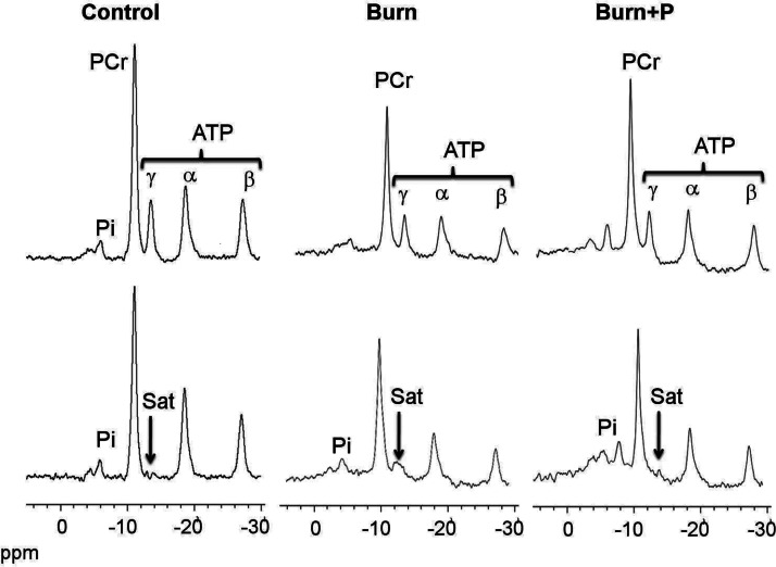
Figure 1.
NMR spectra from 31P NMR saturation-transfer on the hindlimb skeletal muscle of live mice. Representative summed 31P NMR spectra acquired from control, burned (burn), and burned after SS-31 peptide injection (burn+P) mice before (top spectra) and after (bottom spectra) saturation of the γ-ATP resonance. Arrow indicates the position of saturation (Sat) by RF irradiation (−13.2 ppm, γ-ATP). ppm, chemical shift in parts per million.

