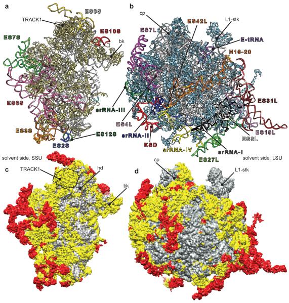
Fig. 2.
Atomic model of the T. brucei ribosome. (a) SSU and (b) LSU: atomic models with the ESs colored and annotated similarly as in Fig. 1. (c) SSU and (d) LSU: atomic models in surface presentations. Gray regions indicate the location of conserved common core elements of all ribosomes, prokaryotic and eukaryotic. Yellow regions highlight eukaryotic-specific conserved elements, including those for trypanosomes. Red regions indicate trypanosome-specific elements, nonexistent in other 80S ribosomes.

