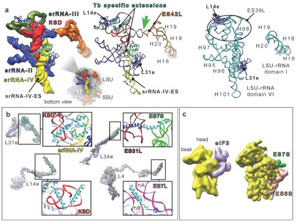
Fig. 4.
T. brucei srRNAs and protein extensions. (a) Left and middle, srRNAs II to IV and KSD cryo-EM density segmentations along with their atomic models compared to yeast domain VI (right). Thick Green arrow highlights the kissing-loop interaction. Black circle surrounds ES39L in yeast ribosome. (b) Ribosomal proteins presenting specific extensions in T. brucei (cyan ribbon) with zooms on the interactions of their extensions with the surrounding rRNA. (c) Left, cryo-EM-derived model of the eIF3-binding site (recreated according to ref. 21). Right, segmented map of the SSU, from T. brucei ribosome, filtered to 20Å. KSD = kinetoplastid-specific domain.

