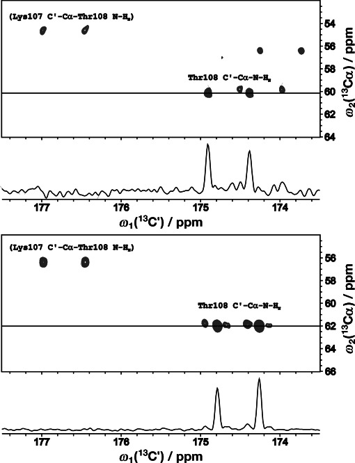
Figure 1.

F1 (C′)–F2 (Cα) planes from the 4D HNCACO-CCR(C′/NHN) spectrum of cBASP1 at pH 2 (top) and pH 6 (bottom) showing JNH-resolved doublets for threonine 108 residue. Labels for interresidual cross-peaks (irrelevant here) are given in parentheses. Noteworthy is the significant difference in relative intensity of the doublet lines.
