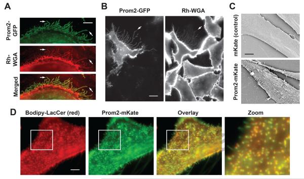
Fig. 1. Prom2 expression induced extensive protrusions that colocalize with a lipid raft marker.
HSFs (A) or CHO cells (B) were transfected with Prom2 GFP (48 hr) and stained with Rh-WGA to label cell surface carbohydrates. Note the extensive protrusions labeled with both Prom2-GFP and Rh-WGA (e.g., at arrows) Bar, 10 μm. (C) HSFs were transfected with Prom2-mKate or mKate only and SEM was performed on fixed cells. Transfected cells showed extensive protrusions on their surface whereas control cells had very few protrusions. Bar, 5 μm. (D) Cells transfected with Prom2-mKate, were incubated with Bodipy-LacCer and fluorescence images of living cells were acquired for Prom-2-mKate and Bodipy-LacCer and merged. Note the extensive co-localization of Prom2 with Bodipy-LacCer. Imaging at low temperature inhibited endocytosis.

