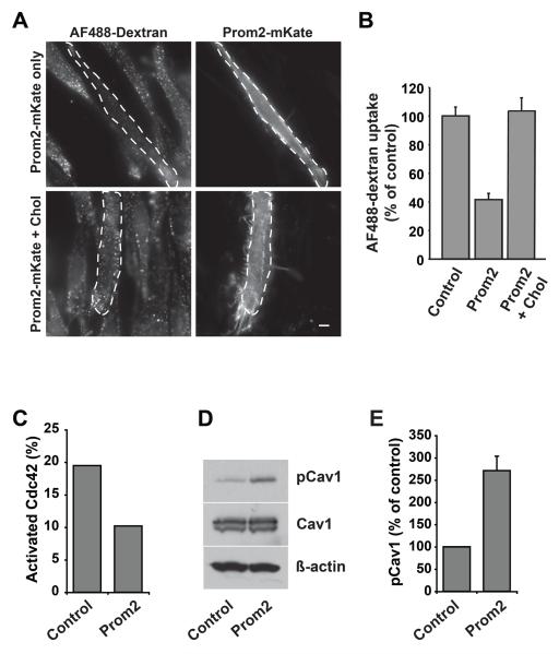
Fig. 3. Prom2 expression decreased fluid-phase endocytosis, decreased Cdc42 activation and stimulated Y14-phosphorylation of caveolin-1.
HSFs transfected ± Prom2-mKate (48 h) were assayed for dextran (fluid phase uptake) endocytosis. In some cases transfected cells were pretreated with 5mM MßCD/Chol for 30 min at 37°C before endocytosis. (A) Fluorescence images of endocytosis of dextran. Transfected cells are outlined by dashed lines. Bars, 10 μm. Dextran endocytosis was quantified (B) by image analysis as in Fig. 2B. Values are means ± SE (n >30 cells in three independent experiments). Note the inhibition of dextran uptake in Prom2-transfected cells and restoration of dextran endocytosis in Prom2-transfected cells by cholesterol. (C) Cdc42 activation assay was performed in cell lysates from transfected and non-transfected cells. Activated Cdc42 (Cdc42-GTP) and total Cdc42 were immunoblotted and levels of activated Cdc42 were normalized to total Cdc42. Note the decreased activation of Cdc42 in Prom2-transfected cells. (D) Cell lysates (as above) were immunoblotted for Y14-pCav1, Cav1 and actin. Note the increased Cav1 phosphorylation in Prom2-transfected cells compared to mkate alone transfected cells. (E) Quantitation of Y14-pCav1 immunoblots. Values are means ± SE (control 100%) for three different experiments.

